About surgery
Heart disease in animals is classified into two types: congenital heart disease in which abnormalities exist since birth in the heart or blood vessels, and acquired heart disease that develops after birth. Both types of heart disease require adequate tests, diagnosis, and treatment.
For humans, heart disease can be treated by surgery using an artificial heart-lung apparatus in many cases. Conversely for animals, the number of institutions and veterinarians that can conduct heart surgery is so low that cardiology care with drugs is provided in most cases. However, there are many cases where heart disease can be cured successfully only after surgery.
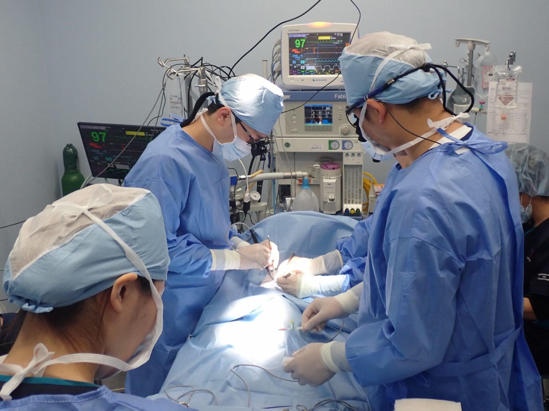
We have introduced extracorporeal circulation devices and special medical devices, which are necessary to perform cardiac surgery, and organized team staff who have accumulated experience in cardiac surgery, so that we are performing cardiac surgery aiming at a radical cure for heart disease. For families who are worried about heart disease in pet dogs or cats, please feel free to consult us.
Indications for surgery
Congenital heart disease: Pulmonary stenosis, atrial septal defects, ventricular septal defects, patent ductus arteriosus
Acquired heart disease: Mitral regurgitation, tricuspid valve incompetence, pacemaker implantation for arrhythmia, cardiac tumors, etc.
About surgery technique
Mitral regurgitation (MR)
The mitral valve, which lies between the left atrium and left ventricle, functions to prevent the backflow of blood, keeping the blood flowing in one direction. Mitral regurgitation is a condition in which the mitral valve has a problem impeding the one-way flow of blood, resulting in symptoms of heart failure.
Mitral regurgitation is the most common heart disease in dogs, which represents the largest number of cases in our hospital.
Cardiology care can only reduce or slow the progression of symptoms. On the other hand, surgery can eliminate heart disease findings or discontinue or reduce drugs.
For mitral regurgitation, we provide mitral valvuloplasty (MVP), which transforms the abnormal mitral valve to the normal state under extracorporeal circulation.
Atrial septal defect (ASD)/ventricular septal defect (VSD)
An atrial septal defect is a congenital heart disease in which there is a congenital hole since birth in the wall that divides the upper area of the heart into left and right sides (left and right atria). In a ventricular septal defect, the hole is in the wall that divides the lower area of the heart into left and right sides (left and right ventricles).
When the hole is very small, it cannot cause a problem even without being treated; however, if the size of the hole is large enough to potentially cause heart failure, surgery to repair the hole needs to be performed at the earliest possible stage.
For an atrial septal defect and a ventricular septal defect, we perform catheter interventional therapy or surgery that repairs a hole directly by using an extracorporeal circulation device.
Pacemaker implantation for arrhythmia (atrioventricular block)
Atrioventricular block, a form of arrhythmia, is a condition where the heart rate is lowered, causing fainting and staggering.
It is often misdiagnosed as a brain disease or other disease.
Diagnosis is confirmed by electrocardiogram or Holter monitoring, which records the electrical activity of the heart for long periods of time in daily life. To treat it, pacemaker implantation is necessary as with a human.
Pulmonary stenosis (PS)
It is a congenital heart disease in which the pulmonary valve (or its adjacent area) in the pulmonary arteries (carry blood from the heart to the lungs) becomes narrow, resulting in impeding blood flow from the heart to the lungs.
Cases of non-severe stenosis might need no treatment or only cardiology care, whereas severe stenosis requires surgery.
Pulmonary stenosis can be treated with balloon dilatation to widen the stricture during catheterization. If catheterization is difficult, open-heart surgery is required.
Patent ductus arteriosus (PDA)
The ductus arteriosus is a small blood vessel that exists in the fetus and connects between the aorta and the pulmonary artery.
The ductus arteriosus usually closes rapidly after birth. PDA is a congenital heart disease in which the vessel remains open even after the baby’s development. If the ductus arteriosus remains open after birth, it can stress the heart, which would progress to heart failure.
Treatment for patent ductus arteriosus requires surgery. The disease can be completely cured by catheter interventional therapy or surgery.
Cardiac tumors
In dogs, it is relatively common for tumors to develop in the heart itself or around it.
Tumors developing in the heart, or around it, include angiosarcoma, aortic body tumors (chemodectoma), ectopic thyroid cancer, and other tumors.Depending on the type and size of the tumors, excision by surgery or incision of the pericardium surrounding the heart can prolong survival or relieve symptoms.